
- वर्तमान स्थान:होम पेज >> स्वास्थ्य संबंधी जानकारी
什么是影像学检查?
2026-06-30 16:07:28
### 影像学检查概述
Imaging examination is a medical diagnostic method that uses various imaging technologies to observe the internal structure and function of the human body, mainly including X-ray, CT, MRI, ultrasound and nuclear medicine. Its core value lies in obtaining lesion information non-invasively or minimally invasively to assist disease screening, diagnosis and treatment evaluation. तकनीकी सिद्धांतों के अनुसार इसे विभाजित किया जा सकता है电离辐射类(如X线,CT)औरएमआरआई जांच परीक्षण (एमआरआई जांच),临床应用需权衡精准度与安全性。
影像学检查的核心流程包含Patient preparation, equipment adjustment, image acquisition and report interpretationचार लिंक. उदाहरण के लिए, सीटी जांच के लिए 4 घंटे से अधिक समय तक भूखे रहने की आवश्यकता होती है, और एमआरआई के लिए धातु की वस्तुओं को हटाने की आवश्यकता होती है। In terms of equipment, GE Healthcare, Siemens, and Philips are the mainstream manufacturers, with representative products such as GE Revolution CT and Siemens MAGNETOM MRI. The examination results are usually comprehensively analyzed by radiologists in combination with clinical history to avoid misjudgment of a single imaging conclusion.
| जांच प्रकार | सिद्धांत | लागू परिदृश्य |
|---|---|---|
| एक्स-रे | आयनकारी विकिरण प्रवेश इमेजिंग | फ्रैक्चर और निमोनिया की जांच |
| सीटी | 多角度X线断层扫描 | ट्यूमर और रक्तस्राव का निदान |
| एमआरआई | 磁场激发氢原子共振 | 神经系统,软组织病变 |
एक्स-रे परीक्षा एक काली और सफेद कंट्रास्ट छवि बनाने के लिए मानव ऊतकों की किरणों की विभिन्न अवशोषण दरों का उपयोग करती है। उच्च घनत्व वाले ऊतक जैसे हड्डियाँ सफेद दिखाई देती हैं, और हवा युक्त संरचनाएँ जैसे फेफड़े काले दिखाई देते हैं। Modern digital DR (direct digital radiography) equipment such as the United Imaging uDR 780i reduces the dose of traditional film by 30%. However, pregnant women and children should use it with caution and use lead protection if necessary.
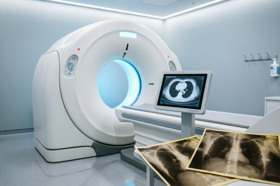
CT obtains tomographic images through X-ray tube rotation scanning, and spiral CT with more than 64 layers can perform three-dimensional reconstruction. Taking lung cancer screening as an example, low-dose CT can detect nodules larger than 2 mm, which is five times more sensitive than chest X-ray (according to 2023 data from the Chinese Journal of Radiology). However, the radiation dose of a single chest CT is about 5-8mSv, which is equivalent to 2-3 years of natural background radiation, and the indications need to be strictly controlled.
### 特殊检查注意事项एमआरआई एक मजबूत चुंबकीय क्षेत्र में हाइड्रोजन परमाणुओं के संकेत परिवर्तनों की इमेजिंग पर निर्भर करता है। इसमें आयनकारी विकिरण नहीं है लेकिन जांच का समय लंबा (आमतौर पर 30-60 मिनट) है। क्लौस्ट्रफ़ोबिया वाले मरीज़ सीमेंस मैग्नेटोम फ्री.मैक्स जैसे खुले एमआरआई उपकरण चुन सकते हैं। विशेष ध्यान देने की आवश्यकता है: पेसमेकर और कॉक्लियर प्रत्यारोपण जैसे धातु प्रत्यारोपण उपकरण विफलता या मानव चोट का कारण बन सकते हैं। सर्जरी से पहले एक सुरक्षा निरीक्षण प्रश्नावली पूरी की जानी चाहिए।
Ultrasound examination uses high-frequency sound waves for real-time imaging, which is suitable for dynamic observation such as fetal development (GE Voluson E10 four-dimensional ultrasound) and heart valve movement. इसके फायदे शून्य विकिरण और दोहराए जाने योग्य ऑपरेशन हैं, लेकिन यह ऑपरेटर के अनुभव पर अत्यधिक निर्भर है। इंटरवेंशनल अल्ट्रासाउंड 1 मिमी तक की सटीकता के साथ सुई बायोप्सी का मार्गदर्शन भी कर सकता है, जो पारंपरिक सर्जरी के जोखिमों को काफी कम कर देता है।
### 综合应用与发展趋势आधुनिक इमेजिंग रुझानमल्टीमॉडल फ्यूजनFor example, PET-CT combines functional metabolism and anatomical imaging to achieve an accuracy of more than 90% in tumor staging (quoted from "Technical Specifications for Nuclear Medicine"). Artificial intelligence-assisted diagnosis systems such as InferRead CT pulmonary nodule detection by InferMedical can improve the reading efficiency by 30%. भविष्य में, आणविक इमेजिंग से बीमारियों के अति-प्रारंभिक दृश्य को सक्षम करने की उम्मीद है।
选择检查方式需综合考虑疾病特征,辐射防护,经济成本तीन तत्व. उदाहरण के लिए, तीव्र पेट के लिए अल्ट्रासाउंड या सीटी को प्राथमिकता दी जाती है, और न्यूरोलॉजिकल घावों के लिए एमआरआई को प्राथमिकता दी जाती है। Patients should truthfully inform their medical history and allergy history, and retain imaging data after the examination for follow-up and comparison. Medical institutions need to regularly calibrate equipment to ensure that imaging quality meets the standards of the "Radiation Diagnosis and Treatment Management Regulations".
उद्धरण स्रोत:
1. "Technical Guidelines for Medical Imaging Examinations" 2022 Edition by the Radiology Branch of the Chinese Medical Association
2. राष्ट्रीय स्वास्थ्य आयोग का "विकिरण निदान और उपचार प्रबंधन विनियम" 2021 संशोधित संस्करण
3. उपकरण डेटा जीई हेल्थकेयर और सीमेंस हेल्थकेयर की आधिकारिक वेबसाइटों पर बताए गए तकनीकी मापदंडों से आता है।
प्रासंगिक ज्ञान
- 2026-10-01महिलाएं वैज्ञानिक तरीके से अपनी त्वचा की देखभाल कैसे करती हैं?
- 2026-10-01नारी सौंदर्य क्या है?
- 2026-09-30महिलाएं वैज्ञानिक तरीके से कैल्शियम की पूर्ति कैसे कर सकती हैं?
- 2026-09-30कैल्शियम अनुपूरण क्या है?
- 2026-09-30अस्थि घनत्व की जांच कितनी बार करानी चाहिए?
- 2026-09-29ऑस्टियोपोरोसिस को कैसे रोकें?
- 2026-09-29महिलाओं को ऑस्टियोपोरोसिस होने का खतरा क्यों होता है?
- 2026-09-29ऑस्टियोपोरोसिस क्या है?
- 2026-09-28क्या हार्मोन रिप्लेसमेंट थेरेपी सुरक्षित है?
- 2026-09-28हार्मोन रिप्लेसमेंट थेरेपी क्या है?
- 2026-09-28रजोनिवृत्ति का प्रबंधन कैसे करें?
- 2026-09-28रजोनिवृत्ति के लक्षण क्या हैं?
- 2026-09-28रजोनिवृत्ति क्या है?
- 2026-09-28प्रसवोत्तर समीक्षा में क्या शामिल है?
- 2026-09-27प्रसवोत्तर समीक्षा क्या है?
- 2026-09-27क्या पेल्विक फ्लोर की मांसपेशियों की मरम्मत आवश्यक है?
- 2026-09-27प्रसवोत्तर पेल्विक फ्लोर मांसपेशियों की मरम्मत क्या है?
- 2026-09-27प्रसव के बाद वैज्ञानिक तरीके से वजन कैसे कम करें?
चीनी औषधीय सामग्री
अधिक-
देखें
बैंगनी मोतियों की प्रभावकारिता और कार्य
बैंगनी मोतियों की प्रभावकारिता और कार्यों का अवलोकनपुरपुरिया औषधीय महत्व वाला एक पौधा है। इसकी पत्तियों, जड़ों और फलों का उपयोग औषधि के रूप में किया जा सकता है। इसके मुख्य कार्यों में शामिल
-
देखें
टिड्डी बीन की प्रभावकारिता और कार्य
टिड्डी बीन की प्रभावकारिता और कार्यों का अवलोकनसोफोरा बीन, जिसे सोफोरा जपोनिका या सोफोरा जपोनिका के नाम से भी जाना जाता है, सोफोरा जपोनिका पेड़ का परिपक्व फल है, जो एक फलीदार पौधा है, और पारंपर
-
देखें
बैजिरेन की प्रभावकारिता और कार्य
बैजिरेन की प्रभावकारिता और कार्यों का अवलोकनबैजिरेन कप्रेसेसी परिवार के एक पौधे प्लैटाइक्लाडस ओरिएंटलिस का सूखा और परिपक्व बीज गिरी है। पारंपरिक चीनी चिकित्सा का मानना है कि यह प्रकृति
-
देखें
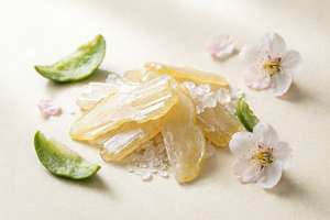
बेर के स्लाइस की प्रभावकारिता और कार्य
बेर के स्लाइस के प्रभाव और कार्यों का अवलोकनबेर के टुकड़े, जिसे बोर्नियोल या डिप्टरोकार्प के नाम से भी जाना जाता है, एक पारंपरिक चीनी औषधीय सामग्री है जिसमें दिमाग को फिर से जीवंत करने, गर्मी क
-
देखें
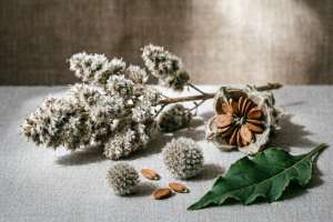
कोचिया छाल की प्रभावकारिता और कार्य
कोचिया छाल की प्रभावकारिता और कार्यों का अवलोकनकोचिया छाल, जिसे झाड़ू अंकुर के रूप में भी जाना जाता है, पारंपरिक चीनी चिकित्सा में गर्मी को दूर करने और नमी को बढ़ावा देने के लिए आमतौर पर इस्ते
-
देखें
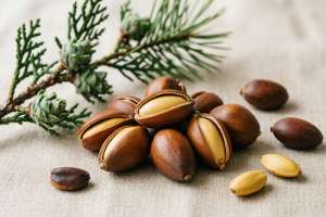
बैजिरेन की प्रभावकारिता और कार्य
बैजिरेन की प्रभावकारिता और कार्यों का अवलोकनबैजिरेन कप्रेसेसी परिवार के एक पौधे, प्लैटाइक्लाडस ओरिएंटलिस का सूखा और परिपक्व बीज गिरी है। पारंपरिक चीनी चिकित्सा का मानना है कि यह प्रकृति
-
देखें
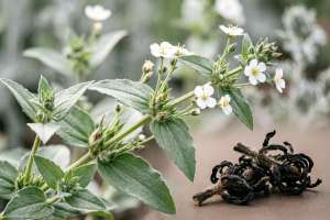
एक्लिप्टा एक्लिप्टा प्रभावकारिता और कार्य
एक्लिप्टा की प्रभावकारिता और कार्यों का अवलोकनएक्लिप्टा, जिसे एक्लिप्टा या एक्लिप्टा भी कहा जाता है, पारंपरिक चीनी चिकित्सा में लीवर और किडनी को पोषण देने, रक्त को ठंडा करने और रक्तस्राव को
-

देखें
हिप्पोकैम्पस का कार्य और भूमिका
हिप्पोकैम्पस के कार्यों और कार्यों का अवलोकनसीहॉर्स एक बहुमूल्य समुद्री जीव है जिसे पारंपरिक चिकित्सा और आधुनिक अनुसंधान दोनों में इसके कई लाभों के लिए पहचाना जाता है।इसके मुख्य कार्यों म
-
देखें
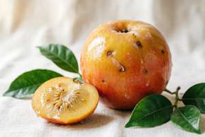
सुगंधित फल के प्रभाव एवं कार्य
सुगंधित फल के प्रभाव और कार्यों का अवलोकनज़ियांगगुओ, जिसे लिगस्टिकम चुआनक्सिओनग या शन्नाई के नाम से भी जाना जाता है, औषधीय और खाद्य दोनों महत्व वाला एक पौधा है, और इसके प्रकंदों का उपयोग अक्सर
-
देखें
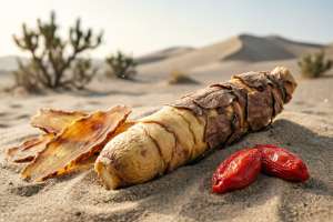
सिस्टैंच डेजर्टिकोला की प्रभावकारिता और कार्य
सिस्टैंच डेजर्टिकोला की प्रभावकारिता और कार्यों का अवलोकनसिस्टैंच डेजर्टिकोला, जिसे सिस्टैंच डेजर्टिकोला भी कहा जाता है, एक पारंपरिक चीनी औषधीय सामग्री है जो मुख्य रूप से रेगिस्तानी क्षे
-
देखें
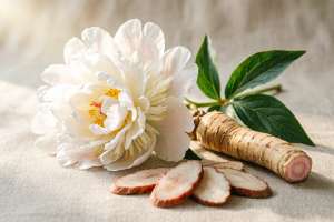
सफेद चपरासी का प्रभाव और कार्य
सफ़ेद पेनी जड़ की प्रभावकारिता और कार्यों का अवलोकनसफेद चपरासी की जड़ पारंपरिक चीनी चिकित्सा में आमतौर पर इस्तेमाल की जाने वाली रक्त-पौष्टिक और मासिक धर्म-नियमन वाली दवा है।रक्त को पोषण देत
-
देखें
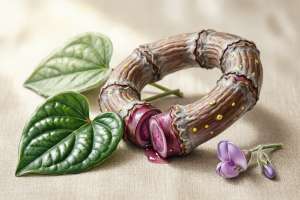
वू हुआ ज़ू टेंग की प्रभावकारिता और कार्य
स्पैथोलोबस की प्रभावकारिता और कार्यों का अवलोकनपांच फूलों वाली ब्लडवाइन एक पारंपरिक चीनी औषधीय सामग्री है, जो मुख्य रूप से हैरक्त परिसंचरण को बढ़ावा देता है और रक्त ठहराव को दूर करता है, दर्